Receiving your 20-week anomaly scan report can feel like trying to read a foreign language, filled with measurements and technical terms. The core problem is the gap between the clinical data on the page and what it actually means for your baby’s health. This guide, written from a senior sonographer’s perspective, translates that jargon into plain English. We’ll explain not just *what* we measure but *why*, demystifying the process so you can feel informed and confident in your antenatal care journey.
You have the letter in your hand. The one from the hospital with the anomaly scan results. It’s a page of numbers, acronyms, and medical terms: BPD, HC, AC, FL, ‘Echogenic Bowel’. You were told at the appointment that everything looked fine, but the printed report feels dense and intimidating. Well-meaning advice often tells you “not to worry if the sonographer is quiet” or that “most scans are normal,” but that doesn’t help you decipher what ‘femur length: 32mm’ actually signifies. You might even wonder if you should have booked one of those private 4D scans for a “better look.”
As a senior sonographer within the NHS, I see this uncertainty every day. The truth is, the anomaly scan isn’t just about taking pictures; it’s a systematic conversation we have with your baby’s developing anatomy. The silence in the room isn’t a sign of trouble; it’s a sign of intense concentration as we meticulously follow a clinical checklist. The measurements aren’t arbitrary; they are pieces of a puzzle that, when put together, give us a detailed assessment of your baby’s wellbeing.
But what if the key to understanding your report wasn’t about memorising medical definitions, but about understanding the sonographer’s perspective? This guide aims to pull back the curtain on the ultrasound room. We’ll move beyond the jargon to explain the clinical reasoning behind the checks we perform. We’ll translate what those measurements mean, why some findings are less concerning than they sound, and how the entire NHS screening pathway is designed to support you and your baby.
This article will walk you through the key milestones of your NHS ultrasound journey. We’ll start by clarifying the timing and purpose of each major scan and test. Then, we will dive into the specifics of what we’re looking at, from the early dating scan to the detailed 20-week assessment, and explain how to prepare to help us get the clearest possible images. We will also tackle common sources of anxiety, such as specific scan findings and the debate between different measurement techniques, before concluding with how to effectively communicate your preferences to your maternity team.
Contents: Decoding Your Pregnancy Scan Journey
- When to Book Your Dating Scan, Anomaly Scan, and Glucose Test in the NHS System?
- Why the Dating Scan Measures the Baby’s Head and Not the Whole Body?
- How to Prepare for Your 20-Week Scan So You Actually See Your Baby Clearly?
- Echogenic Bowel on Your Scan: Why 95% of Cases Mean Nothing Serious?
- The £150 4D Bonding Scan That Gives You Pretty Pictures but Zero Medical Information
- Ultrasound Growth Estimate vs Fundal Height: Which Predicts Birth Weight Better?
- When Your Midwife Should Refer You for Extra Growth Scans in the Third Trimester?
- Why Your Birth Plan Should Fit on One Page for Busy Midwives to Actually Read?
When to Book Your Dating Scan, Anomaly Scan, and Glucose Test in the NHS System?
Navigating the NHS antenatal pathway can feel like a complex schedule of appointments. However, each test is timed with precise clinical reasoning to give us the most accurate information at the most effective time. Missing these windows can sometimes reduce the effectiveness of the screening. Understanding this timeline is the first step to feeling in control of your pregnancy care journey.
The dating scan, for instance, is optimally performed between 11 and 14 weeks. This is the most accurate time to establish your due date and is also the critical window for the combined screening test for Down’s, Edwards’ and Patau’s syndromes. The 20-week anomaly scan is timed for a point when the baby’s organs are large enough to be seen clearly, but there is still sufficient time for any necessary follow-up or decisions. Finally, the Glucose Tolerance Test (GTT) for gestational diabetes is scheduled between 24 and 28 weeks, as this is when pregnancy-related insulin resistance typically peaks. The table below, based on the standard NHS antenatal screening timeline, breaks down the purpose and timing of these key appointments.
| Scan/Test | Timing (Weeks) | Clinical Purpose | What Happens If You Miss the Window |
|---|---|---|---|
| Dating Scan (with optional NT screening) | 8-14 weeks (ideally 11-13+6) | Establish accurate due date (±5-7 days), confirm viability, detect multiple pregnancy, combined screening for Down’s/Edwards/Patau’s syndromes | Quad test offered 14-20 weeks (less accurate). Due date estimated from 20-week scan if no earlier scan performed |
| 20-Week Anomaly Scan | 18-20+6 weeks (up to 23 weeks in some cases) | Screen for 11 physical conditions, check organ development when visible but time remains for decision-making, assess placental position and amniotic fluid | Reduced detection rates for some conditions. Incomplete screening must be documented. No further routine scan offered |
| Glucose Tolerance Test (GTT) | 24-28 weeks | Screen for gestational diabetes at peak insulin resistance period, enabling dietary/medication management before third trimester | Later diagnosis reduces time for blood sugar management. May miss optimal window for preventing macrosomia complications |
| Growth Scans (if indicated) | 28+ weeks (third trimester) | Monitor fetal growth when risk factors present (previous SGA/LGA, diabetes, hypertension, reduced movements), assess wellbeing via Doppler | Only offered when clinical indication exists – not routine for all pregnancies. Minimum 3-week intervals between scans |
Ultimately, these appointments are a structured offer of care, not a mandatory obligation. While you have the right to decline any screening, understanding the purpose and timing of each test empowers you to make an informed decision that feels right for you.
Why the Dating Scan Measures the Baby’s Head and Not the Whole Body?
At your first scan, often called the dating scan, it can be surprising that we focus so intently on measuring your baby from head to bottom, known as the Crown-Rump Length (CRL). It seems logical to measure the whole baby, including the legs, but in early pregnancy, this method is by far the most accurate for establishing your estimated due date (EDD).
The reason for this is consistency. In the first trimester, all foetuses grow at a remarkably predictable rate. The CRL measurement is less affected by individual genetic variations than other parameters that appear later. From about 7 to 13 weeks, there is a direct and reliable correlation between this length and gestational age. The legs are not included because they are often curled up and difficult to measure accurately, which would introduce significant variability. After 14 weeks, the baby begins to curl and uncurl more, making CRL less reliable. At this point, we switch to a combination of head circumference (HC), biparietal diameter (BPD), abdominal circumference (AC), and femur length (FL) to assess growth.
This early precision is crucial. As clinical guidance from the American College of Obstetricians and Gynecologists confirms, there is an accuracy of just ±5-7 days for CRL measurements taken before 14 weeks. This accurate EDD becomes the baseline for your entire pregnancy, ensuring that all subsequent growth assessments and screening tests, like the 20-week anomaly scan, are performed at the correct time. An inaccurate due date could lead to misinterpretation of growth later on, causing unnecessary worry or missed opportunities for intervention.
So, when we focus on that tiny measurement, we are not ignoring the rest of your baby; we are establishing the single most important data point for monitoring the health and progress of your entire pregnancy.
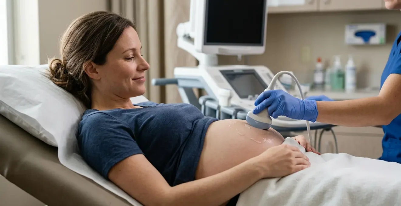
How to Prepare for Your 20-Week Scan So You Actually See Your Baby Clearly?
The 20-week anomaly scan is a detailed anatomical survey. To get the clear, high-quality images needed to check everything from the baby’s heart to their kidneys, we need your help. How you prepare for the appointment can make a significant difference to the quality of the scan and can even mean the difference between a completed check and needing to come back for a second appointment.
The single most important preparation step is arriving with a comfortably full bladder. Drinking about a pint (500ml) of water an hour beforehand is ideal. This isn’t to make you uncomfortable; a full bladder acts as an ‘acoustic window’. It lifts the uterus out of the pelvis and pushes the bowel, which can be full of image-disrupting gas, out of the way. This gives us a much clearer, unobstructed view of your baby. Another key factor is your baby’s position. If your baby is in a difficult position (for example, lying with their spine towards us), it can be impossible to get a good view of the heart. We might ask you to go for a walk, jiggle your bump, or have a cold drink to encourage the baby to move.
The entire examination takes, on average, approximately 30 minutes, but it can take longer if the baby is uncooperative. It’s also important to prepare mentally. We conduct the scan in a darkened room to see the screen better, and there will be periods of silence. This is when we are concentrating, taking complex measurements, and methodically working through our clinical checklist. It’s not a sign that something is wrong. Knowing this beforehand can help you and your partner feel more relaxed during the process.
By working together, you help us perform the most thorough check possible. Your preparation allows us to focus entirely on the clinical dialogue with your baby’s anatomy, ensuring we gather all the information needed to confirm their wellbeing.
Echogenic Bowel on Your Scan: Why 95% of Cases Mean Nothing Serious?
Of all the technical terms on a scan report, ‘echogenic bowel’ is one that often causes immediate alarm. It simply means the baby’s bowel appears brighter than usual on the ultrasound, as bright as the surrounding bone. When parents hear this, their minds can race to the worst-case scenarios they find online, like cystic fibrosis or chromosomal abnormalities. While these are associations, it’s crucial to understand that in the vast majority of cases, an echogenic bowel is what we call an isolated benign variant.
This finding is relatively uncommon, with research indicating that echogenic bowel is seen in only about 0.2% to 1.8% of second-trimester scans. When it is an ‘isolated’ finding—meaning every other part of the baby’s anatomy survey is completely normal, and your first-trimester screening results were low-risk—the outcome is overwhelmingly positive. It can be caused by the baby swallowing a small amount of blood from a minor bleed earlier in the pregnancy, something that is completely harmless and resolves on its own. It can also simply be a normal variation with no underlying cause.
However, because of the small association with other conditions, we take it seriously. If we see an echogenic bowel, standard NHS protocol is to refer you to a Fetal Medicine specialist. This is not a reason to panic; it is a precaution. The specialist will likely recommend further testing, which may include a detailed scan to re-examine the baby’s anatomy, blood tests for infections like cytomegalovirus (CMV), and a test for cystic fibrosis. It is this thorough follow-up that allows us to rule out other issues and, in most cases, provide complete reassurance that it is a transient, harmless finding. The key is to see it not as a diagnosis, but as a reason for a closer look.
The pathway is designed to be cautious. We refer for further checks to be absolutely certain, but the statistics and clinical experience show that for an isolated echogenic bowel, a healthy baby is by far the most likely outcome.
The £150 4D Bonding Scan That Gives You Pretty Pictures but Zero Medical Information
The rise of private “boutique” scan clinics offering 4D “bonding” or “reassurance” scans can create confusion. For around £150, you get beautiful, moving 3D images of your baby’s face, a longer appointment, and a relaxed, family-friendly atmosphere. It’s a wonderful experience, but it is crucial to understand the fundamental difference between this and your NHS anomaly scan: one is entertainment imaging, and the other is a diagnostic medical examination.
A 4D bonding scan is optimised for aesthetics. The sonographer’s goal is to get a clear, cute picture of the baby’s face. They are not typically performing the systematic, 11-point anatomical check that is the entire purpose of the 20-week NHS scan. The person performing the scan may not even be a qualified diagnostic sonographer registered with the Health and Care Professions Council (HCPC). Their expertise might be in operating the 3D/4D software, not in identifying complex cardiac defects or measuring amniotic fluid levels. A “reassurance” scan that shows you a heartbeat and a cute wave does not reassure you about the baby’s kidney function or brain development.
This doesn’t mean private scans have no value, but their value is emotional, not medical. Problems arise when parents either skip their NHS scan in favour of a private one, or when a private clinic oversteps its remit and either misses a problem or raises a concern without a proper NHS referral pathway in place. A qualified diagnostic sonographer in an NHS setting is trained to follow strict protocols (like those from the British Medical Ultrasound Society – BMUS) and has an integrated system for immediate referral to Fetal Medicine if a problem is suspected. Before you book a private scan, it is vital to arm yourself with the right questions to ensure you are making an informed choice about your and your baby’s health.
Your Action Plan: Key Questions for a Private Scan Clinic
- Is the person performing the scan a qualified diagnostic sonographer registered with the Health and Care Professions Council (HCPC)?
- Will a detailed medical report be provided, or is this purely for images and bonding purposes?
- What specific conditions or abnormalities is the scan designed to detect, if any?
- If an abnormality is suspected, what is the referral pathway and will results be shared with my NHS care team?
- Does the clinic follow BMUS (British Medical Ultrasound Society) clinical guidelines for fetal scanning?
- What is the clinic’s policy on scanning duration – is there adequate time for diagnostic assessment or just entertainment imaging?
- Are there additional costs for follow-up consultations if findings need clarification?
- Does the scan replace or supplement my NHS anomaly scan – and does the clinic recommend I still attend my NHS appointment?
Think of it this way: your NHS scan is the essential structural survey of the house, while the 4D scan is the beautifully shot promotional video. Both have their place, but only one tells you if the foundations are sound.
Ultrasound Growth Estimate vs Fundal Height: Which Predicts Birth Weight Better?
From around 24 weeks, your midwife will start measuring your “fundal height”—the distance from your pubic bone to the top of your uterus—at every appointment. If this measurement deviates from your personalised growth chart, you’ll be referred for a growth scan. This can cause a lot of anxiety, but it’s important to understand the different roles these two methods play. Neither is perfect, but they work together as a screening and diagnostic partnership.
Fundal height is a simple, non-invasive screening tool. It’s like a quick check to see if everything is roughly on track. Its main strength is in tracking a trend over time on your baby’s own personalised GROW (Gestation Related Optimal Weight) chart. A single measurement is not very informative, but a pattern of measurements that flattens out or suddenly jumps up is a flag that tells us we need a closer look. It is a low-cost, routine way to pick up potential issues.
An ultrasound growth scan is the more detailed diagnostic tool we use when that flag is raised. We take multiple measurements—head, abdomen, and femur—and use a complex algorithm to calculate an Estimated Fetal Weight (EFW). This is more precise than a tape measure on your bump, but it is still an estimate. The baby’s position, the amount of amniotic fluid, and maternal body shape can all affect the measurements. In fact, comprehensive clinical research shows a margin of error of ±15% between the estimated weight on a scan and the baby’s actual birth weight. This means a baby estimated at 7lb 7oz (3.4kg) could be born anywhere between 6lb 6oz and 8lb 8oz.
So, which is better? The answer is that they are not in competition. Fundal height is the sensitive smoke detector, and the ultrasound scan is the detailed inspection by the fire brigade. One alerts us to a potential issue, and the other gives us the detailed information needed to make a clinical plan.
When Your Midwife Should Refer You for Extra Growth Scans in the Third Trimester?
In a low-risk pregnancy, the 20-week anomaly scan is often the last routine ultrasound you’ll have. Further “growth scans” are only performed in the third trimester if there is a specific clinical reason to monitor the baby’s growth or wellbeing more closely. It’s your midwife’s job, using fundal height measurements and assessing your overall health, to identify the red flags that warrant this referral.
The most common trigger is a deviation in fundal height measurement. This isn’t about your bump being “too big” or “too small” compared to others; it’s about how your baby is growing against their own personalised growth curve on their GROW chart. According to NHS guidance, a referral is typically made if the measurement plots below the 10th centile or above the 97th centile, or if the growth pattern appears to slow down or plateau over several appointments. This indicates the baby may be small for gestational age (SGA) or large for gestational age (LGA), both of which may require closer monitoring.
However, fundal height is not the only trigger. A midwife will also refer you for a growth scan for a number of other important reasons that relate to maternal or fetal health. These scans do more than just estimate weight; they also assess the amount of amniotic fluid and check the blood flow through the umbilical cord (Doppler studies) to assess placental function. The following are all standard reasons for a referral:
- Maternal high blood pressure or the development of pre-eclampsia.
- A diagnosis of gestational diabetes, which can lead to a large baby (macrosomia).
- A significant reduction or change in the baby’s movement patterns as reported by you.
- A previous pregnancy that resulted in an SGA or LGA baby.
- Carrying twins or more, which requires regular growth monitoring for all babies.
- Other maternal health conditions, like kidney disease, that could affect placental function.
A referral for a growth scan is not automatically a cause for alarm. It is a proactive and precautionary step to get a more detailed picture, ensuring that if any support is needed for you or your baby, it can be provided in a timely manner.
Key takeaways
- Your NHS anomaly scan is a detailed medical examination, not an aesthetic “photo shoot”; the sonographer’s silence is often a sign of deep concentration.
- Scan preparation, especially having a full bladder, creates an “acoustic window” that is critical for helping the sonographer get clear images.
- Unexpected findings like “echogenic bowel” are often benign variations, and the referral for specialist review is a precautionary step, not a definitive diagnosis of a problem.
Why Your Birth Plan Should Fit on One Page for Busy Midwives to Actually Read?
After spending months decoding clinical reports, the final piece of communication is translating your own wishes to the delivery team. This is the purpose of a birth plan. However, in the fast-paced environment of a labour ward, a multi-page, highly detailed document is simply not practical. For a birth plan to be an effective communication tool, it needs to be a concise, one-page summary that a busy midwife can absorb and reference at a glance.
Think of it as a “medical passport” for your labour, not a prescriptive script. The goal is to give the team, who may be meeting you for the first time, a quick and clear understanding of your key preferences, fears, and medical background. A well-written one-page plan shows that you have thought about your options while also understanding that labour can be unpredictable. It fosters a collaborative atmosphere, positioning your preferences as a starting point for a conversation, not a rigid list of demands. A midwife is far more likely to engage positively with a clear, respectful summary than an overwhelming essay.
The most effective birth plans focus on the essentials. They highlight what is most important to you, acknowledge flexibility, and provide critical information that will help the team support you better. According to guidance from the NHS, the key elements to include are your birth partner’s details, your pain relief preferences, any significant fears (like needle phobia), and your initial feeding intentions. By keeping it to one page, you make it a genuinely useful tool for the people who will be by your side.
- Birth partner details: Full name and contact number.
- Pain relief preferences: A clear indication (e.g., ‘prefer water/movement first’, ‘epidural from outset’) while acknowledging flexibility.
- Any significant fears or previous trauma: A brief note so the team can provide appropriate support.
- Delivery position preferences: Note if you want freedom to move or use a birthing pool, while staying open to clinical needs.
- Immediate post-birth preferences: Skin-to-skin contact, delayed cord clamping, Vitamin K preference.
- Feeding intentions: Breastfeeding, formula, or undecided.
- Special circumstances: Any clinical factors like gestational diabetes or Group B Strep status.
Having navigated the complexities of antenatal screening, you are now equipped to communicate effectively with your maternity team. Use your birth plan not as a contract, but as the start of your final and most important conversation of your pregnancy, ensuring your preferences are understood and respected as you welcome your baby.